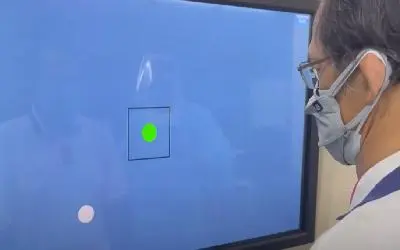
Recent technological advancements have enabled researchers to transition from 2D to Opti-Speech technology, presenting 3D images of the tongue. This innovation, credited to the University of Texas at Dallas, forms the basis of a new study suggesting that observing 3D tongue movements aids in learning speech sounds.
Dr. William Katz, a co-author of the study and a professor at UT Dallas’ Callier Center for Communication Disorders, noted the potential significance of these findings for stroke patients seeking to enhance their speech articulation.
“Observing 3D tongue images can contribute to teaching consonant sounds,” explained Katz, who teaches at UT Dallas School of Behavioral and Brain Sciences. “Our goal is not only to teach consonant sounds through visual aids but also to comprehend apraxia and related disorders by utilizing visual feedback.”
Published in the journal Frontiers in Human Neuroscience, the study, though small-scale, demonstrated that exposure to visual feedback training enhanced participants’ accuracy in learning new sounds.
Katz is among the pioneers proposing that visual feedback on tongue movements could aid stroke patients in speech recovery.
“Those with apraxia of speech often struggle with this process. Despite knowing what they intend to say, they encounter difficulty in executing their speech plans, resulting in incorrect sounds,” Katz elaborated.
Initially motivated to showcase patients their tongues, Katz emphasized how this visual demonstration could reveal the correct articulation of sounds.
The recent technological leap to Opti-Speech technology from 2D visuals allows for displaying 3D images of the tongue. Earlier UT Dallas research confirmed that the Opti-Speech visual feedback system effectively provides real-time feedback for speech learning.
The study delved into compensatory articulation, wherein rapid acoustic shifts lead subjects to believe they are producing one sound while hearing feedback indicating another.
“People immediately readjust their articulation when the sound pushes them in a certain direction. Then, upon turning off the shift, they overcompensate,” explained Katz. “In our study, we visually shifted participants’ tongue movements gradually. As a result, they adjusted their sounds to match the tongue image.”
Katz underscored the importance of visualizing body movements in rehabilitation therapy, acknowledging the necessity for further exploration.
“Our aim is to unravel why visual feedback impacts speech. Is it primarily compensatory or does it involve mirroring or entrainment? Do individuals guide their tongue visually, relying then on a sense of ‘mouth feel’? Which brain regions are likely involved?” Katz pondered.
“3D imaging is forging a new path for speech rehabilitation. Hopefully, this research will soon aid patients striving to improve their speech.”
Note: Material may have been edited for length and content. For further information, please contact the cited source.
Publication
Katz WF, Mehta S Visual Feedback of Tongue Movement for Novel Speech Sound Learning. Frontiers in Human Neuroscience, Published November 19 2015. doi: 10.3389/fnhum.2015.00612